Cultures
Dermatophytest
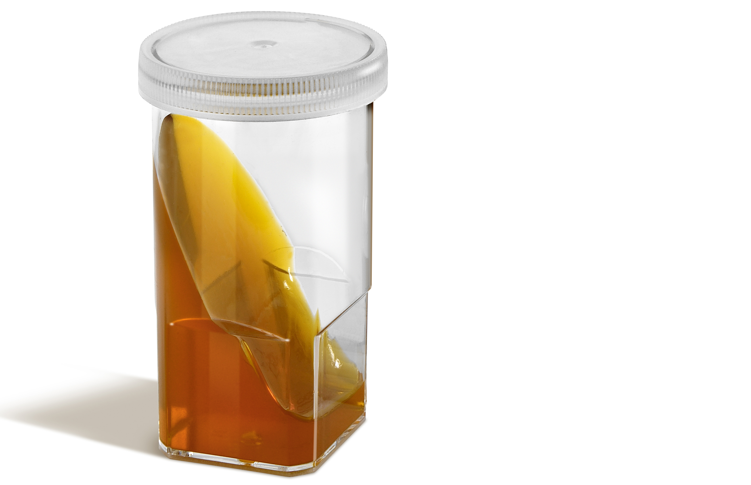
Dermatophytes-specific culture medium for selective growth of Trichophyton and Microsporum in pets.
Dermatophytest is a dermatophyte-selective medium for the specific growth of the genus Microsporum canis, Microsporum gypseum, Microsporum persicolor and Tricophyton mentagrophytes.
- Dermatophytest makes it possible to identify the fungal agents responsible for dermatomycosis (ringworm) in different species of
![180002_Kit_Speed-Dermatophytest_x1_right.png]() pets (cats, dogs, guinea pigs, rabbits, mice, etc.).
pets (cats, dogs, guinea pigs, rabbits, mice, etc.). - Interpretation is eased by a change in color of the agar indicating growth of dermatophytes within 2-14 days.
- Colonies growing on the agar surface can be easily picked and observed under a microscope for more accurate species identification.
- Selective compounds in the agar help limit the growth of unwanted contaminants such as other fungi or bacteria.
For veterinary use only.
Product specifications
| Sample | Skin scratches, skin swab, scales, hairs, calws, feathers |
|---|---|
| Species | Dogs, Cats, roddents, rabbits... |
| Result reading | From day 2 to day 14 |
| Storage | Store between +2°C and +8°C, away from light |
| Presentation | Box of 6 vials |
Why using Dermatophytest ?
Dermatomycoses, such as ringworm, are due to the proliferation of different fungal agents of the genus Microsporum and Trichophyton. This dermatological infection is very frequent in cats, dogs, rodents or lagomorphs.
Transmission to humans is possible, in particular with Microsporum canis or Trichophyton Mentagrophytes, the most common agents responsible for Ringworm. These agents are closed as agents of zoonosis (2).
The spores, disseminated by infected animals in the environment, reach keratinized areas (hair follicles, stratum corneum, claws), form hyphae and generate lesions which can develop into chronicity.
Some animals are healthy carriers but can contaminate their peers or owners. It is important to check any lesion compatible with dermatomycosis in order to implement sanitary measures quickly and avoid spread.
The culture test is the most reliable method to diagnose dermatophytes, unlike direct examination under a microscope which requires experience and lacks sensitivity (3). The skin infected areas to be sampled can be facilitated by a Wood's lamp examination.
When to use Dermatophytest ?
Dermatophytest is indicated :
-
As soon as a clinical suspicion exists: localized or multifocal alopecic lesions, crusty or scaly dermatosis, seborrhea, claw deformation, etc., culture is recommended.
-
To assess the need of anti-fungal therapy in the case of surinfected lesions
-
To identify asymptomatic or transient mechanical carrier animals in order to prevent zoonotic risk to susceptible individuals (4).
The diagnosis of ringworm must combine the results of the various additional examinations carried out by the veterinarian.
Quick guide
For complete instuctions, please refer to the Instruction For Use of the products.
- Sampling
Wash skin lesions with water and soap (no antifungal nor disinfectant solutions) or with 70° alcohol and dry it.
- Collect hairs and scales by brushing or plucking the edge of a lesion using sterile tweezers. Gently place the hairs and scales on the agar.
- By scraping cut: using the edge of a glass blade or the back of a scalpel blade, scrape starting from the center of the infected area to the healthy part of the skin. Gently place the hairs and scales on the agar.
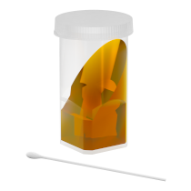
- By swabbing : first moisten the end of a sterile swab with a few drops of sterile distilled water. Pass the swab over the entire lesion and discharge the swab onto the agar using alternating movements, without crushing the agar.
2. Culture
-
Leave the cap slighlty open. Culture needs air exchange to grow.
-
Incubate at room temperature, or on a thermostat around 27°, away from direct light.
3. Reading the result
Start checking any growth or color change from day 2 and every 2 days until day 14 if necessary.
References
(1) CARUSO K.J. et coll. Skin scraping from a cat [dermatophytosis]. Vet Clin. Pathol, 2002,31,13-15
(2) CARLOTTI D.N., PIN D. Aspects cliniques, histopathologiques, diagnostic différentiel et traitement des dermatomycoses canines et félines. Ann Med Vet, 2002, 146, 85-96
(3) MORIELLO K.A. Practical diagnostic testing for dermatophytosis in cats. Vet Med, 2003, 98, 859-875 MIGNON B et coll. Le portage asymptomatique de Microsporum canis chez le chat. Annales de Médecine Vétérinaire, 2001, 145, 174-177
(4) MIGNON B et coll. Le portage asymptomatique de Microsporum canis chez le chat. Annales de Médecine Vétérinaire, 2001, 145, 174-177.
Websites
EUROPEAN SCIENTIFIC COUNSEL COMPANION ANIMAL PARASITES (ESCCAP)


 pets (cats, dogs, guinea pigs, rabbits, mice, etc.).